

Computed tomography (CT) is a medical imaging technique used in radiology to get detailed images of the body non-invasively. CT scanners use a rotating x-ray tub and a row of detectors placed in the gantry to measure x-ray attenuations by different tissues inside the body. The multiple x-ray measurements taken from different angles are then processed on a computer using reconstruction algorithms to produce cross-sectional slices of the body.
Contrast enhanced CT (CE-CT) can also be used to visualise structures within the body which would otherwise be difficult to delineate from their surroundings. Contrast agents are usually iodine based.

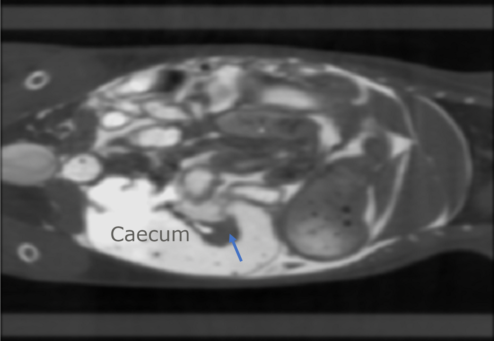
CT Image of mouse abdomen

CT image of mouse lungs. High levels of air in lungs allows contrast against surrounding tissue
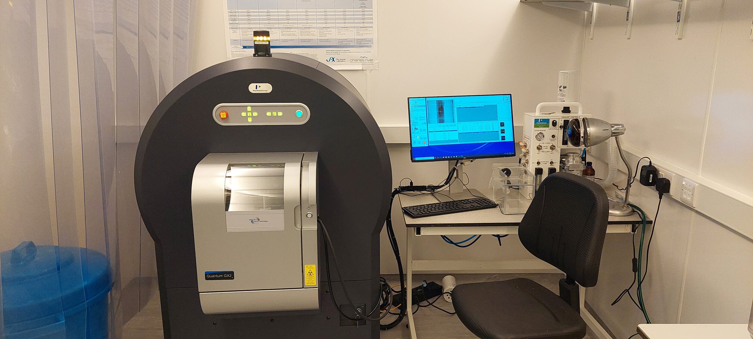
Within NPIC we utilise the Quantum GX CT (Perkin Elmer)
CECT image of mouse abdomen. Negatively contrasted tumour (Indicated by arrow) is visible against highly contrasted abdominal cavity.